Hello Everyone, It’s still your favourite Medic Writer and I will like to welcome us all into todays session. Today we will be discussing a major landmark in the abdominal region which is highly important for diagnosis, treatment and a major landmark during surgical operations. The abdominal region has a the highest number of viserals which are important for body metabolism.
Today we will be examining the INGUINAL CANAL.
The inguinal canal is a short passage that extends inferiorly and medially through the inferior part of the abdominal wall. It is superior and parallel to the inguinal ligament.
The canal serves as a pathway by which structures can pass from the abdominal wall to the external genitalia. It is of clinical importance as a potential weakness in the abdominal wall, and thus a common site of herniation.
In summary of that, the inguinal canal can also be liken to a normal fossa e.g Cubital, poptileal etc and every fossa must have a boundary and must also contain materials i.e contents.
The BOUNDARIES OF THE INGUINAL CANAL
The inguinal canal is bordered by anterior, posterior, superior (roof) and inferior (floor) walls. It has two openings – the superficial and deep rings.
Walls
Anterior wall – aponeurosis of the external oblique, reinforced by the internal oblique muscle laterally.
Posterior wall – transversalis fascia.
Roof – transversalis fascia, internal oblique, and transversus abdominis
.
Floor – inguinal ligament (a ‘rolled up’ portion of the external oblique
aponeurosis), thickened medially by the lacunar ligament.
The contents of the inguinal canal includes the;
The spermatic cord in males only
The round ligament in females only
The illioingunial Nerve
The genital branch of genitofemoral Nerve
Thank you all for following; You can get back to us on the comment box or through medicspen2022@gmail.com
Medics_Writer✍️✍️✍️
The Inguinal canal
Hello Everyone, It’s still your favourite Medic Writer and I will like to welcome us all into todays session. Today we will be discussing a major landmark in the abdominal region which is highly important for diagnosis, treatment and a major landmark during surgical operations. The abdominal region has a the highest number of viserals which are important for body metabolism.
Today we will be examining the INGUINAL CANAL.
The inguinal canal is a short passage that extends inferiorly and medially through the inferior part of the abdominal wall. It is superior and parallel to the inguinal ligament.
The canal serves as a pathway by which structures can pass from the abdominal wall to the external genitalia. It is of clinical importance as a potential weakness in the abdominal wall, and thus a common site of herniation.
In summary of that, the inguinal canal can also be liken to a normal fossa e.g Cubital, poptileal etc and every fossa must have a boundary and must also contain materials i.e contents.
The BOUNDARIES OF THE INGUINAL CANAL
The inguinal canal is bordered by anterior, posterior, superior (roof) and inferior (floor) walls. It has two openings – the superficial and deep rings.
Walls
Anterior wall – aponeurosis of the external oblique, reinforced by the internal oblique muscle laterally.
Posterior wall – transversalis fascia.
Roof – transversalis fascia, internal oblique, and transversus abdominis
.
Floor – inguinal ligament (a ‘rolled up’ portion of the external oblique
aponeurosis), thickened medially by the lacunar ligament.
The contents of the inguinal canal includes the;
The spermatic cord in males only
The round ligament in females only
The illioingunial Nerve
The genital branch of genitofemoral Nerve
Thank you all for following; You can get back to us on the comment box or through medicspen2022@gmail.com
Medics_Writer✍️✍️✍️
The Peritoneum
Hi everyone, it’s still your favourite Medic Writer and today we will discussing a very important anatomical structures which is highly responsible for spread of disease, infection, nutrients and other anatomical materials in the abdominal region.
It most be noted that the abdomen is one of the most complicated regions of the body and his majorly due to the large amount of organs to the body and their relevancy to the body metabolism.
So very quick we will be discussing about the PERITONEUM, which we all know to be a membranous lining that covers the abdominal cavity and the abdominal viserals.
The peritoneum is a continuous membrane which lines the abdominal cavity and covers the abdominal organs (abdominal viscera).
It acts to support the viscera, and provides pathways for blood vessels and lymph to travel to and from the viscera. From the definition above we can all deduce that the peritoneum covers majorly all the abdomen and can be divided into two main types which are,
PARIETAL PERITONEUM
The parietal peritoneum lines the internal surface of the abdominopelvic wall. It is derived from somatic mesoderm in the embryo.
It receives the same somatic nerve supply as the region of the abdominal wall that it lines; therefore, pain from the parietal peritoneum is well localised. Parietal peritoneum is sensitive to pressure, pain, laceration and temperature.
VISCERA PERITONEUM
The visceral peritoneum invaginates to cover the majority of the abdominal viscera. It is derived from splanchnic mesoderm in the embryo.
The visceral peritoneum has the same autonomic nerve supply as the viscera it covers. Unlike the parietal peritoneum, pain from the visceral peritoneum is poorly localised and the visceral peritoneum is only sensitive to stretch and chemical irritation.
Thanks all for following me, please note that your comments, suggestions are all important to us, you can also get back to us at
medicspen2022@gmail.com.
Medics_Pen✍️
The Anterior Abdominal wall
Hi everyone, still your favourite medic writer and today I will be bringing to us this very interesting anatomical clinical correlate. Will be discussing majorly today about the ABDOMINAL WALL
The abdominal wall consist of the skin located superficially, the underlying connective tissue, the muscles and other anatomical structures which will be discussed during the course of the class, You can always give us feedback in the comment space or via our email
medicspen2022@gmail.com……
The Abdominal wall encloses the abdominal cavity, i.e the abdominal wall serves as a blanket covering the viserals of the abdomen, the abdominal cavity is just the space of the abdomen containing viserals and other anatomical structures.
Today majorly will be discussing on the clinical correlate of the abdominal wall as said earlier, the clinical correlate will focus on INCISION, incision simply means a cut to create an entrance to a particular part or region.
The major site of incision in the abdominal wall is the LINEA ALBA and it’s due to its low vascularization
An incision that is made through the linea alba. It can be extended the whole length of the abdomen by curving around the umbilicus. The linea alba is poorly vascularised, so blood loss is minimal, and major nerves are avoided. It can be used in any procedure that requires access to the abdominal cavity.
Paramedian
Similar to the median incision, but is performed laterally to the linea alba, providing access to more lateral structures (kidney, spleen and adrenals). This method ligates the blood and nerve supply to muscles medial to the incision, resulting in their atrophy.
Kocher
A Kocher incision ③ begins inferior to the xiphoid process and extends inferolaterally in parallel to the right costal margin. It is mainly used to gain access for gall bladder and/or biliary tree pathology.
Two modifications and extensions of the Kocher incision are possible:
Chevron / rooftop incision or modification ④ – the extension of the incision to the other side of the abdomen. This may be used for oesophagectomy, gastrectomy, bilateral adrenalectomy, hepatic resections, or liver transplantation
Mercedes Benz incision or modification ⑤ – the Chevron incision with a vertical incision and break through the xiphisternum. This may be used for the same indications as the Chevron incision, however classically seen in liver transplantation.
McBurney
A McBurney is a called a ‘grid iron’ incision, because it consists of two perpendicular lines, splitting the fibres of the muscles without cutting them – this allows for excellent healing. McBurney incision is performed at McBurney’s point (1/3 of the distance between the ASIS and the umbilicus) and is used in an open appendicectomy. Thanks everyone for reading. Don’t forget to make use of the comme
THE CUBITAL FOSSA
THE CUBITAL FOSSA
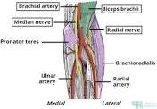
The cubital fossa is a triangular depression which marks the area of transition between the arm and the forearm. The cubital fossa is located above the elbow joint and located anteriorly in a balanced anatomical position.
The cubital fossa is an area of transition between the anatomical arm and the forearm. It is located as a depression on the anterior surface of the elbow joint.
BORDERS OF THE CUBITAL FOSSA
The cubital fossa is triangular in shape, and thus has three borders, mainly bounded by the muscles of the arm and forearm.
Lateral border – medial border of the brachioradialis muscle.
Medial border – lateral border of the pronator teres muscle.
Superior border – hypothetical line between the epicondyles of the humerus.
The floor of the cubital fossa is formed proximally by the brachialis, and distally by the supinator muscle. The roof consists of skin and fascia, and is reinforced by the bicipital aponeurosis. Within the roof runs the median cubital vein, which can be accessed for venepuncture.
CONTENTS OF THE CUBITAL FOSSA
The cubital fossa is mainly filled with fats.
prominent contents include;
The contents of the cubital fossa include vessels, nerves and the biceps tendon (lateral to medial):
Radial nerve – this is not always strictly considered part of the cubital fossa, but is in the vicinity, passing underneath the brachioradialis muscle. As it does so, the radial nerve divides into its deep and superficial branches.
Biceps tendon – runs through the cubital fossa, attaching to the radial tuberosity, just distal to the neck of the radius.
Brachial artery – supplies oxygenated blood to the forearm. It bifurcates into the radial and ulnar arteries at the apex of the cubital fossa.
Median nerve – leaves the cubital between the two heads of the pronator teres. It supplies the majority of the flexor muscles in the forearm.
CLINICAL CORRELATE
Venepuncture
The median cubital vein is located superficially within the roof of the cubital fossa. It connects the basilic and cephalic veins, and can be accessed easily – this makes it a common site for venepuncture.
THE BILE
Bile is an aqueous, alkaline, greenish-yellow liquid whose main function is to emulsify fats in the small intestine and to eliminate substances from the liver. The liver produces 0.25-1L of bile per day.
In this article, we shall consider the many different components of bile, and how these are produced in the liver
THE CONSTITUENTS OF BILE
Bile is made up of bile acids, cholesterol, phospholipids, bile pigments (such as bilirubin and biliverdin), electrolytes and water.
These are split into two main groups, known as the bile acid-dependent and bile acid-independent components:
The bile acid-dependent component is produced by hepatocytes. The hepatocytes secrete bile acids, bile pigments and cholesterol into canaliculi, which are small channels that transport the bile acid dependent portion towards the bile ducts.
The bile acid-independent component is made by the ductal cells that line the bile ducts. These cells secrete an alkaline solution – similar to the fluid made by pancreatic duct cells. The hormone secretin stimulates this secretion.
Both the dependent and independent components of bile enter the intrahepatic bile ducts which drain into the biliary tree, a series of ducts which transport bile from the liver to the gallbladder and duodenum.
Bile is continuously produced, but we only need it during and after meals. Therefore the gallbladder concentrates and stores bile, by removing the water and ions. After eating, the hormone cholecystokinin is released from the duodenum. This stimulates gallbladder contraction and relaxes the sphincter of Oddi, thus allowing bile to flow into the duodenum.
THE BILE PIGMENT
Bile also contains bile pigments which are excretory products of the liver and include biliverdin and bilirubin. Bilirubin is a breakdown product of haemoglobin and is conjugated in the liver and secreted into bile. Bile pigments are normally excreted in the faeces and make the faeces appear brown. Problems in the liver or biliary tree often result in the accumulation of bilirubin in the blood, which manifests as jaundice.
Enterohepatic Circulation of Bile Acids
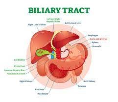
Bile passes out of the liver through the bile ducts and is concentrated and stored in the gallbladder. During and after a meal, bile is excreted from the gall bladder by contraction and passes into the duodenum through the common bile duct. Most of the bile acids are reabsorbed in the terminal ileum and returned to the liver via the hepatic portal vein. The liver then extracts the bile salts.
The enterohepatic circulation allows the liver to recycle and preserve a pool of bile acids.
The colon
COLON
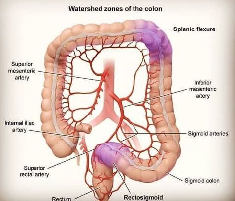
The colon is one of the most important organ that helps in digestion, the colon is like a stomach pipe that helps in transportation and absorption of food nutrients including food particles. Colon also helps in the secretion of some materials that helps in digestion.
The colon is also known as the large bowel or large intestine. It is an organ that is part of the digestive system (also called the digestive tract) in the human body. The digestive system is the group of organs that allow us to eat and to use the food we eat to fuel our bodies.
Food begins in the mouth where it is chewed by the teeth into smaller pieces. Once swallowed the food travels into the esophagus which connects to the stomach.
In the stomach food is further broken down to liquid and passed on to the small bowel (intestine).
In the small bowel, the food breakdown continues with the assistance of the pancreas, liver and gallbladder. Here is where all the important vitamins and nutrients in food are absorbed.
What is left over, which is mostly liquid, then moves into the colon. The water is absorbed in the colon. Bacteria in the colon break down the remaining material. Then the colon moves the leftover material into the rectum.
The rectum is like a storage-holder for this waste. Muscles in the rectum move the waste, called stool, out of the body through the anus.
medics_ writer’s
Breast cancer
Good day guys, I will be sharing with us the causes, diagnosis, and few treatment of breast cancer.
Breast cancer is one of the most common disease in our present world, According to research, breast cancer is one of the disorder causing the highest death rate in our world.
Scientifically, cancer is a disease in which some of the body’s cells grow uncontrollably and spread to other parts of the body.
Cancer can start almost anywhere in the human body, which is made up of trillions of cells. Normally, human cells grow and multiply (through a process called cell division) to form new cells as the body needs them. When cells grow old or become damaged, they die, and new cells take their place.
DEFINITION OF BREAST CANCER.
In a lame man language, you can define breast cancer as cancer of the tissue containing or involving the milk glands (mammary tissue). But medically can be defined as the cancer that;
Forms in the cells of the breasts. After skin cancer, breast cancer is the most common cancer diagnosed in women in the United States. Breast cancer can occur in both men and women, but it’s far more common in women.
CAUSES OF BREAST CANCER
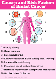
Breast cancer develops as a result of genetic mutations or damage to DNA. These can be associated with exposure to estrogen, inherited genetic defects, or inherited genes that can cause cancer, such as the BRCA1 and BRCA2 genes. When a person is healthy, their immune system attacks any abnormal DNA or growths.
Breast cancer usually starts in the inner lining of the milk ducts or the lobules that supply them with milk. From there, it can spread to other parts of the body.
SIGNS AND SYMPTOMS
Some warning signs of breast cancer are—
New lump in the breast or underarm (armpit).
Thickening or swelling of part of the breast.
Irritation or dimpling of breast skin.
Redness or flaky skin in the nipple area or the breast.
Pulling in of the nipple or pain in the nipple area.
Nipple discharge other than breast milk, including blood.
Any change in the size or the shape of the breast.
Pain in any area of the breast.
Keep in mind that these symptoms can happen with other conditions that are not cancer.
medics_writers
BASIC HOME REMEDIES FOR HIGH BLOOD PRESSURE
Common factors that can lead to high blood pressure include: A diet high in salt, fat, and/or cholesterol. Chronic conditions such as kidney and hormone problems, diabetes, and high cholesterol. Family history, especially if your parents or other close relatives have high blood pressure.
COMMON CAUSES OF BLOOD PRESSURE
Smoking.
Being overweight or obese.
Lack of physical activity.
Too much salt in the diet.
Too much alcohol consumption
Stress.
Older age.
Genetics.
HOME REMEDIES TO PREVENT HIGH BLOOD PRESSURE
1) Regular body exercise
2) Low intake of sodium
3) Consume less alcohol
4) Quite smoking
5) Eat magnesium and calcium rich food
6) Taking deep breath
7) Eat berries
Down Syndrome
Hello guys, today I will be sharing a crucial and very common genetic disorder.
Genetic disorders simply means a mistake in a genetic makeup of an offspring. Genetic disorders are caused by various factors which will be discussed in this article.
DOWN SYNDROMEis a genetic condition that causes mild to serious physical and developmental problems.
People with Down syndrome are born with an extra chromosome. Chromosomes are bundles of genes, and your body relies on having just the right number of them. With Down syndrome, this extra chromosome leads to a range of issues that affect you both mentally and physically.
Down syndrome is a lifelong condition. Although it can’t be cured, doctors know more about it now than ever. If your child has it, getting the right care early on can make a big difference in helping them live a full and meaningful life
And it is very advisable to begin a down syndrome immediately it is noticed or diagnosed because sometimes it is life threatening.
TYPES OF DOWN SYNDROME
There are three types of Down syndrome:
Trisomy 21. This is by far the most common type, where every cell in the body has three copies of chromosome 21 instead of two.
Translocation Down syndrome. In this type, each cell has part of an extra chromosome 21, or an entirely extra one. But it’s attached to another chromosome instead of being on its own.
Mosaic Down syndrome. This is the rarest type, where only some cells have an extra chromosome 21.
you can’t tell what type of Down syndrome someone has just by how they look. The effects of all three types are very similar, but someone with mosaic Down syndrome may not have as many symptoms because fewer cells have the extra chromosome.
SIGNS AND SYMPTOMS OF DOWN SYNDROME
Signs and symptoms of Down syndrome varies in different individuals,
few symptoms include.
Eyes that slant up at the outer corner
Small ears
Flat noses
Protruding tongue
Tiny white spots in the colored part of the eyes
Short neck
Small hands and feet
Short stature
Loose joints
Weak muscle tone
Many people with Down syndrome don’t have any other health issues, but some do. Common conditions include heart problems and trouble hearing and seeing.
There’s no specific treatment for Down syndrome. But there is a wide range of physical and developmental therapies designed to help people with Down syndrome reach their full potential. The earlier you start them, the better. Each child will have different needs. Yours may benefit from:
Physical, occupational, and speech therapy
Specialized education services
Social and recreation activities
Programs that offer job training and teach self-care skills
You’ll also work closely with your child’s doctor to watch for and manage any health problems associated with the condition.